5/15
8:30-11:30am – SY08 PET Scan
- Address: Martinos Center for Biomedical Imaging located in the Charlestown Navy Yard campus of Massachusetts General Hospital (MGH) — 149 13th Street, Charlestown, MA 02129
Multiple System Atrophy (MSA) is a rare and progressive neurodegenerative disorder characterized by a combination of symptoms that affect both the autonomic nervous system and movement. Diagnosing and managing MSA can be challenging due to its complex nature and overlap with other neurodegenerative diseases. One advanced imaging technique that has proven valuable in the assessment of MSA is the SY08 PET scan.
What is a SY08 PET Scan?
https://clinicaltrials.gov/study/NCT06098612
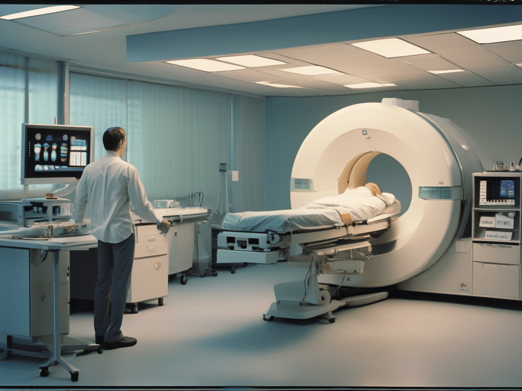
A SY08 PET scan, or Positron Emission Tomography scan, is a sophisticated imaging test that allows doctors to visualize how tissues and organs function within the body. This non-invasive procedure involves the use of a radioactive tracer that highlights areas of high chemical activity, providing detailed, three-dimensional images.
The Role of SY08 PET Scans in MSA:
- Early and Accurate Diagnosis: MSA can be difficult to distinguish from other disorders such as Parkinson’s disease. SY08 PET scans can reveal characteristic patterns of brain activity that help differentiate MSA from other neurodegenerative conditions, enabling a more accurate diagnosis.
- Assessing Brain Function: MSA affects various parts of the brain, including regions responsible for movement, balance, and autonomic functions. PET scans can show abnormalities in these areas by highlighting changes in metabolic activity, offering insights into the extent and progression of the disease.
- Monitoring Disease Progression: SY08 PET scans can track changes in brain function over time, providing valuable information about the progression of MSA. This helps doctors adjust treatment plans as the disease advances, ensuring that patients receive the most appropriate care.
- Evaluating Treatment Effectiveness: By comparing PET scan results before, during, and after treatment, doctors can assess how well a particular therapy is working. This is crucial for managing MSA, as it allows for personalized treatment plans tailored to the individual’s response.
How Does a SY08 PET Scan Work?
- Radioactive Tracer: A small amount of a radioactive substance (tracer) is injected into the bloodstream. This tracer accumulates in areas with high levels of chemical activity, which are often sites of disease.
- Imaging Process: The PET scanner detects gamma rays emitted by the tracer and constructs detailed images of the brain. These images show the metabolic activity of different brain regions.
- Combined Imaging: Often, PET scans are combined with CT or MRI scans to provide more comprehensive images. This combination, known as PET-CT or PET-MRI, enhances diagnostic accuracy.
Benefits of SY08 PET Scans for MSA Patients:
- Detailed Insights: PET scans provide a unique view of brain function, revealing abnormalities that other imaging techniques might miss.
- Early Detection: By identifying changes in brain activity early, PET scans can help diagnose MSA at a stage when intervention might be more effective.
- Tailored Treatments: Understanding the specific brain regions affected by MSA allows doctors to create more targeted and effective treatment plans.
- Improved Quality of Life: Accurate diagnosis and monitoring of MSA can lead to better management of symptoms, potentially improving the quality of life for patients.
In conclusion, SY08 PET scans are a powerful tool in the fight against Multiple System Atrophy. They offer detailed insights into brain function, aiding in early diagnosis, monitoring disease progression, and evaluating treatment effectiveness. For patients with MSA, this advanced imaging technique can make a significant difference in managing their condition and improving their overall quality of life.
~Coach~

Leave a reply to Coach Cancel reply